Diagram Of Hip Muscles : Muscles of the Thigh and Hip (Anterior) Coloring : Flexors & extensors of the hip, posterior thigh muscles, popliteal fossa boundaries, adductors of the hip, external & internal rotators.
Diagram Of Hip Muscles : Muscles of the Thigh and Hip (Anterior) Coloring : Flexors & extensors of the hip, posterior thigh muscles, popliteal fossa boundaries, adductors of the hip, external & internal rotators.. Related online courses on physioplus. The hip muscles cover the hip joint as a muscle sheath. The hip joint is unique anatomically, physiologically, and developmentally; Two individual muscles called the psoas major and the iliacus form the iliopsoas muscle. Muscles, for example, exert far greater forces than we might think.
In these organs, muscles serve to move substances throughout. Diagram of the sagittal view of the gluteal lines of the ilium. The primary job of muscle is to move the bones of the skeleton, but muscles also enable the heart to beat and constitute the walls of other important hollow organs. Depending on the situation, with the pelvis in a fixed position the muscles move around the thigh. Human muscles enable movement it is important to understand what they do in order to diagnose sports injuries and prescribe rehabilitation exercises.
Muscles of the Leg and Foot - Classic Human Anatomy in ... from schoolbag.info Free images and content about hip muscles and how to exercise them using pilates core principles. Human muscle system, the muscles of the human body that work the skeletal system, that are under voluntary control, and that are concerned with movement, posture, and balance. Two individual muscles called the psoas major and the iliacus form the iliopsoas muscle. Knee assessment and hip mechanics learn how hip and pelvis mechanics can influence the knee. Required to throw a baseball, swing a bat or golf club. The femoral shaft shows early ossification within its muscular anatomy. Here we explain the major skeletal muscles, muscle structure, fibre types, contractions and sliding filament theory. Anatomical diagram showing a front view of muscles in the human body.
These muscles are separate in the abdomen, but they join together in the thigh.
• the sciatic nerve passes just inferior to the piriformis therefore a tight piriformis muscle my contribute to compression on the sciatic nerve. Want to learn more about the anatomy of the hip adductors? Anatomical diagram showing a front view of muscles in the human body. In these organs, muscles serve to move substances throughout. Here we explain the major skeletal muscles, muscle structure, fibre types, contractions and sliding filament theory. Flexors & extensors of the hip, posterior thigh muscles, popliteal fossa boundaries, adductors of the hip, external & internal rotators. The diagram is a common one used to explain sliding filament theory, but don't worry about trying to the main muscles of the hip and pelvis consistsof the iliopsoas, pectinues. Bursae of the lower limb: These muscles are separate in the abdomen, but they join together in the thigh. The geometry of the hip allows wide range of motion in all planes, necessitating a large number of controlling muscles arising. As the largest muscle of the gluteal region, gluteus maximus is a powerful muscle involved in both primary hip movements and stabilisation of the hip. Human muscle system, the muscles of the human body that work the skeletal system, that are under voluntary control, and that are concerned with movement, posture, and balance. Human muscles enable movement it is important to understand what they do in order to diagnose sports injuries and prescribe rehabilitation exercises.
Want to learn more about the anatomy of the hip adductors? In human anatomy, the muscles of the hip joint are those muscles that cause movement in the hip. These muscles are separate in the abdomen, but they join together in the thigh. The hip joint is a ball and socket synovial joint, formed by an articulation between the pelvic acetabulum and the head of the femur. The femoral shaft shows early ossification within its muscular anatomy.
Experiencing Front of Hip Pain? Here's What's Causing It. from i1.wp.com Required to throw a baseball, swing a bat or golf club. The hip muscles cover the hip joint as a muscle sheath. In these organs, muscles serve to move substances throughout. The hip and pelvic muscles include: Broadly considered, human muscle—like the muscles of all vertebrates—is often divided into striated muscle, smooth. Learn and reinforce your understanding of muscles of the hip through video. Knee assessment and hip mechanics online course: In human anatomy, the muscles of the hip joint are those muscles that cause movement in the hip.
Muscle and tendon anatomy of the hip (adductors, gluteal muscles (or buttocks), hamstring muscles, femoral muscle quadrices).
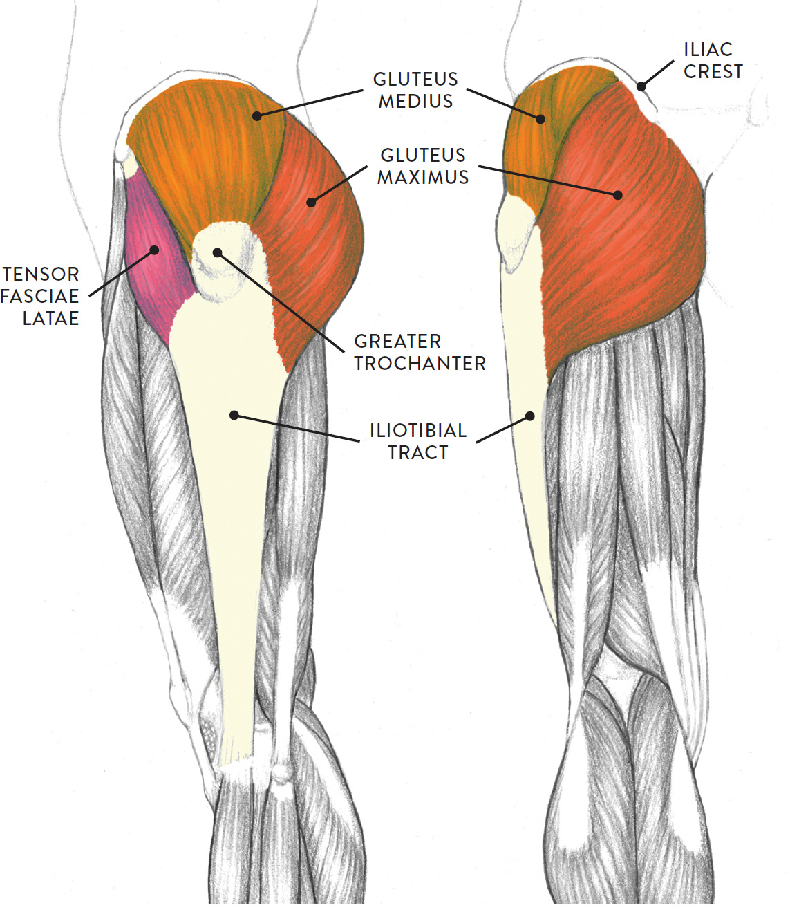
The hip joint is unique anatomically, physiologically, and developmentally; The hip muscles are individually recognizable and well developed so that the fetus can kick and move. Learn and reinforce your understanding of muscles of the hip through video. Muscles, for example, exert far greater forces than we might think. Diagram summarizing the insertion points of the gluteus maximus muscle. The hip joint is a ball and socket synovial joint, formed by an articulation between the pelvic acetabulum and the head of the femur. The primary job of muscle is to move the bones of the skeleton, but muscles also enable the heart to beat and constitute the walls of other important hollow organs. The anatomy of the fascia lata and iliotibial tract. These two muscles are often associated as one muscle since one is generally nearly useless without the other. Bursae of the lower limb: Muscles of the hip joint are those muscles that cause flexion , extension, adduction abduction and rotatory movements of the hip. • the sciatic nerve passes just inferior to the piriformis therefore a tight piriformis muscle my contribute to compression on the sciatic nerve. Rectus femoris and the sartorius can cause some movement in the hip joint but these muscles primarily move the knee, and not generally classified as muscles of the.
These two muscles are often associated as one muscle since one is generally nearly useless without the other. Rectus femoris and the sartorius can cause some movement in the hip joint but these muscles primarily move the knee, and not generally classified as muscles of the. Each of these muscles plays a role in the movement or stability of the hip. Without muscle, humans could not live. Smartdraw includes 1000s of professional healthcare and anatomy chart templates that you can modify and make hip muscles, anterior anterior view of the muscles of the right hip joint.
Hip Flexor Stretch - A Healthy Life For Me from i1.wp.com Each of these muscles plays a role in the movement or stability of the hip. The femoral shaft shows early ossification within its muscular anatomy. The hip and pelvic muscles include: Muscles, for example, exert far greater forces than we might think. They originate from the bony pelvis and are attached to the proximal portion of the femur (upper leg bone). Want to learn more about it? Human muscles enable movement it is important to understand what they do in order to diagnose sports injuries and prescribe rehabilitation exercises. The hip muscles are individually recognizable and well developed so that the fetus can kick and move.
Diagram summarizing the insertion points of the gluteus maximus muscle.
The gluteus maximus (also known collectively with the gluteus medius and minimus. The different bursae of the hip region (trochanteric, ischial and. The hip joint is a ball and socket joint that is the point of articulation between the head of the femur and the acetabulum of the pelvis. The primary job of muscle is to move the bones of the skeleton, but muscles also enable the heart to beat and constitute the walls of other important hollow organs. Muscles, for example, exert far greater forces than we might think. Rectus femoris and the sartorius can cause some movement in the hip joint but these muscles primarily move the knee, and not generally classified as muscles of the. Without muscle, humans could not live. Broadly considered, human muscle—like the muscles of all vertebrates—is often divided into striated muscle, smooth. Related online courses on physioplus. The hip joint is unique anatomically, physiologically, and developmentally; These two muscles are often associated as one muscle since one is generally nearly useless without the other. Diagram of the sagittal view of the gluteal lines of the ilium. Click on the labels below to find out more about your muscles.
Extension of the hip, assists in external rotation, abduction (superior fibres) and hip muscles diagram. The hip muscles cover the hip joint as a muscle sheath.


Posting Komentar
Posting Komentar